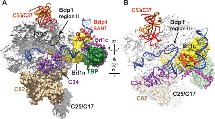
FIG 6.
Positions of Bdp1 region II and the SANT domain in the Pol III open promoter complex model. (A) The model of the Pol III-Brf1-TBP-DNA open promoter complex was initially assembled with the structures of the Pol II-TFIIB-TBP complex, the Brf1-TBP-DNA complex, and Pol I (31, 43–46). The C82 and C34 structures were generated on the basis of homology modeling using structures of human homologues as the templates. The C53/C37 dimerization module was generated using the human TFIIF (Rap74/Rap30) dimer structure as the homology modeling template. C82, C34, and the C53/C37 dimerization module were added to Pol III on the basis of biochemical cross-linking and binding data, as described previously (31). Template and nontemplate DNA strands are in blue and cyan, respectively. The Pol III 12-subunit core structure (white), C82 (tan), the Brf1 cyclin fold repeat domain (yellow), and TBP (dark green) are shown as a molecular surface representation. The magenta sphere in the Pol III core structure represents the active site magnesium ion. C34 (purple), C53 (orange), C37 (red), the Bdp1 SANT domain (orange-red), and Brf1 C-terminal block II (magenta) are shown as backbone trace representations. TFIIB N-terminal zinc ribbon, reader, and linker domains (yellow) are placed in the Pol III active site cleft to indicate likely positions for Brf1 N-terminal zinc ribbon and linker domains. (B) Different orientation of the open promoter complex. Pol III core, C82, and TBP are in semitransparent surface representations.

