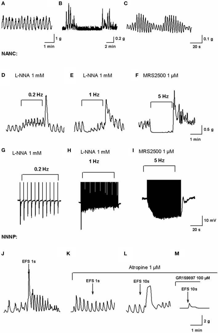
Figure 1.
In vitro motility patterns and enteric neurotransmission in the human colon. Mechanical recording of high frequency (HF) contractions of a constant amplitude probably associated to slow wave activity (A), low-frequency (LF) contractions superimposed to HF contractions (B) and wax and wane of HF contractions amplitude (C) obtained from the experiments performed in our lab (Mane et al., 2014a). Mechanical recording of EFS under NANC conditions on L-NNA 1 mM incubated tissue at a frequency of 0.2 Hz (D) and 1 Hz (E) and MRS2500 1 μM incubated tissue at a frequency of 5 Hz (F) (Mane et al., 2014a). In L-NNA incubated tissue, purinergic neurotransmission is only able to cause phasic relaxations while nitrergic neurotransmission at 5 Hz completely inhibits spontaneous contractions. Electrophysiological recording of electrical field stimulation on L-NNA incubated tissue at a frequency of 0.2 Hz (G) and 1 Hz (H) and MRS2500 incubated tissue at a frequency of 5 Hz (I) (Mane et al., 2014a). Notice how purinergic fast IJP amplitude is reduced with high frequencies of EFS after the first pulse while the nitrergic response increases due to summation of slow IJP. Mechanical recording of EFS in NNNP conditions at 50 Hz, 50 V, 0.4 ms for 1 s (J) eliciting an atropine-sensitive (K) contraction. Mechanical recording of EFS in NNNP conditions at 50 Hz, 50 V, 0.4 ms for 10 s (L) eliciting an antiNK2-sensitive (M) contraction (Martinez-Cutillas et al., 2015).

