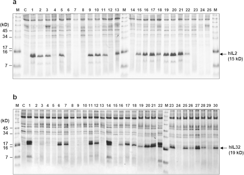
Figure 5. SDS-PAGE analysis of hIL-2 (a) and hIL-32 (b) expressed by randomly selected translational fusion partners from TFP library.
A 0.6-mL aliquot of culture supernatant was precipitated with acetone and analysed on a 10% Tricine gel. M: standard protein size marker, C: host strain carrying a mock vector, (a) Lane 1: TFP1-4, lane 2, 9, 13: TFP5, lane 3: TFP18, lane 5, 22: TFP19-1, lane 10: TFP16-1, lane 11, 15, 18: TFP17-1, lane 16: TFP5-3, lane 17, 19, 20, 21: TFP17-3. (b) Lane 1, 14, 22: TFP10, lane 6, 11, 21, 23: TFP5-1, lane 7: TFP6, lane 12: TFP7-1, lane 16: TFP18-1, lane 17: TFP20, lane 18, 25: TFP5-2, lane 19: TFP11, lane 20: TFP14-1, lane 26: TFP21, lane 27: TFP16-3, lane 28: TFP21, lane 30: TFP22. The protein was revealed by Coomassie staining.

