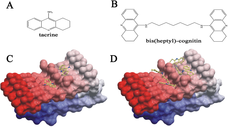
Figure 7. The potential interaction between bis(heptyl)-cognitin and Aβ.
(A) Chemical structure of tacrine. (B) Chemical structure of bis(heptyl)-cognitin. Low-energy binding conformations of bis(heptyl)-cognitin (C) or tacrine (D) bound to the surface of Aβ assemblies (Gly33-Met35 and Met35-Gly37) generated by molecular docking. The small molecule is depicted as a ball-and-stick model showing carbon (yellow), nitrogen (blue), and hydrogen (dark grey) atoms. The Aβ assemblies are shown as skin representation.

