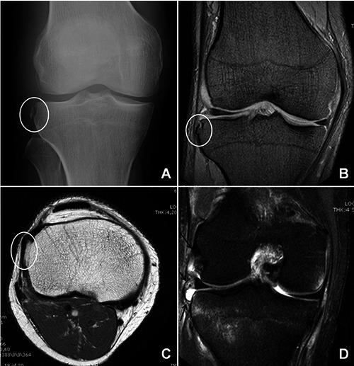
Figure 2.

Imaging. A) Anteroposterior knee radiograph, showing Segond fracture (white circle). B) Coronal magnetic resonance imaging (MRI) showing sequelae of Segond fracture (white circle). C) Sagittal T1 MRI showing sequelae of Segond fracture (white circle). D) Coronal fat suppression MRI showing anterolateral ligament rupture (black arrows).
