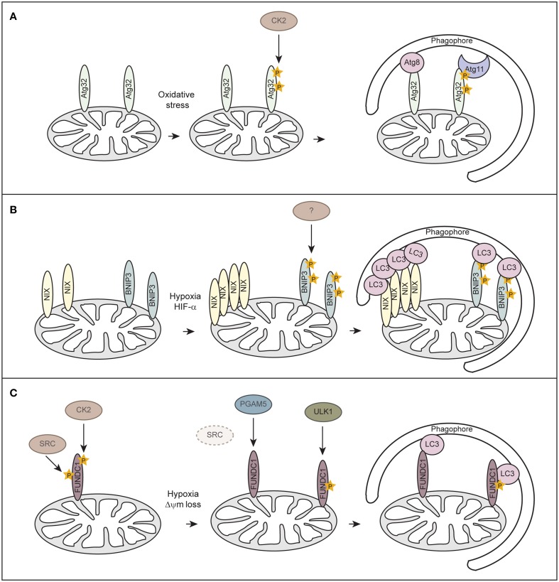
Figure 3.
Receptor-mediated mitophagy. (A) Atg32-mediated mitophagy in S. cerevisiae. Atg32 is an outer mitochondrial membrane protein whose expression is induced upon mitophagy-inducing conditions. Atg32 interacts with Atg8 and Atg11 via distinct domains. Casein kinase 2 (CK2) phosphorylates Atg32 upon mitophagy-inducing conditions, which is essential for the Atg11 interaction without affecting Atg32-Atg8 binding. (B) NIX/BNIP3-mediated mitophagy in mammalian cells. NIX and BNIP3 are outer mitochondrial membrane proteins that interact with LC3 through LIR motifs in their N-terminal region. Upon hypoxia, NIX and BNIP3 are transcriptionally induced in a HIF-α-dependent manner. Phosphorylation of BNIP3 promotes its binding to LC3 and subsequent mitophagy. The kinase for BNIP3 phosphorylation is unknown. (C) FUNDC1-mediated mitophagy in mammalian cells. FUNDC1 is an outer mitochondrial membrane protein that interacts with LC3 through a LIR domain at its cytosol-exposed N-terminus. Under normal physiological conditions, FUNDC1 is phosphorylated by SRC and CK2, thereby preventing LC3 binding. Upon hypoxia or loss of mitochondrial membrane potential (Δψm), the expression of SRC is strongly suppressed and PGAM5 dephosphorylates FUNDC1. Dephosphorylation of FUNDC1 enhances the interaction between FUNDC1 and LC3 and promotes mitophagy. Phosphorylation of FUNDC1 by ULK1 enhances its binding to LC3.

