
Figure 4.
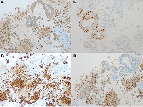
Neoplastic cells (100×) stain positively for thyroid transcription factor-1 (TTF-1) (A). Neoplastic cells (200×) show strong reactivity for calcitonin (B). Neoplastic cells (100×) are negatively stained for estrogen receptor. The adjacent epithelial cells of a mammary duct show estrogen receptor reactivity and is considered as an internal positive control (C). Neoplastic cells (100×) are positively stained for chromogranin (D).