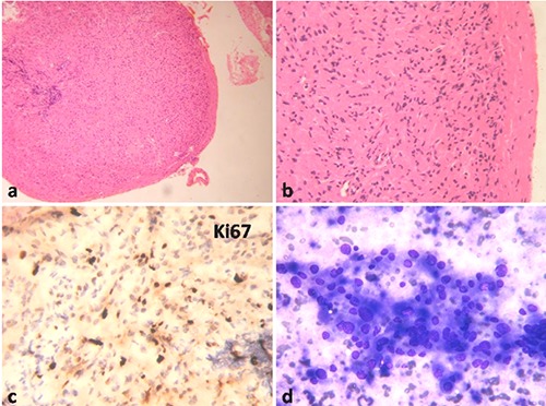
Figure 2.

a) Esophageal tumor nodule at low magnification, Hematoxylin & Eosin (H&E, 100×). b) High magnification showing tumor composed of sheets of polygonal cells with eosinophilic cytoplasm, H&E 400×. c) Low Ki67 proliferative fraction, DAB chromogen 400×. d) Liver aspirate showing loose clusters of polygonal cells with variation in nuclear size and abundant cytoplasm, MGG 400×.
