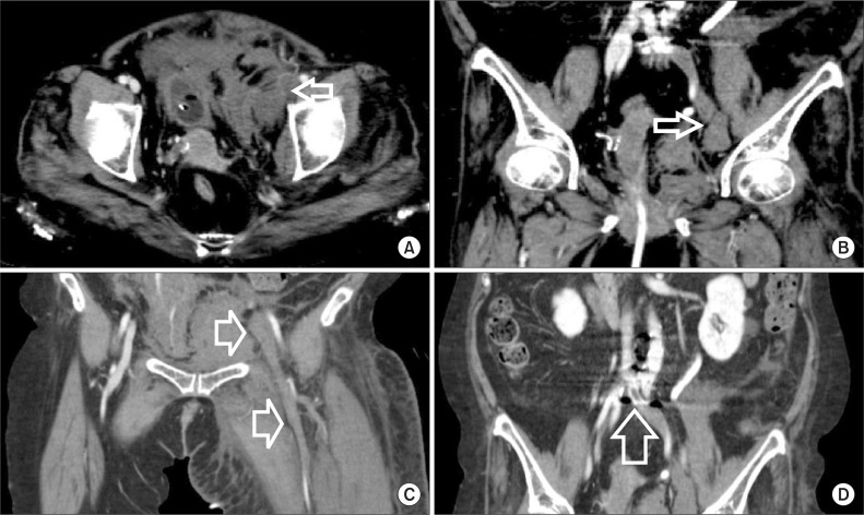
Fig. 1.
Initial abdominopelvic computed tomography showing (A) rupture of the left external iliac vein (EIV) with a hematomoa annotated by arrows. (B) A large hematomoa on the medial side of the left EIV annotated by arrows. (C) Wide-range deep vein thrombosis from left common iliac vein to left popliteal vein. (D) May-Thurner syndrome, overwhelmed iliac vein was annotated.

