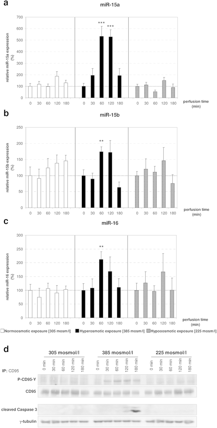
Figure 1. Upregulation of miR-15a/b and miR-16 by hyperosmolarity in perfused rat liver.

Rat livers were perfused with normoosmotic medium (305 mosmol/l), hyperosmotic medium (385 mosmol/l) or hypoosmotic medium (225 mosmol) for up to 180 min. Samples were taken at the time points indicated. (a) miR-15a is significantly upregulated after 60 and 120 minutes of hyperosmotic conditions, whereas a stable expression is observed under hypo- and normoosmotic (305 mosmol/l) conditions (* p < 0.05; **p < 0.01; ***p < 0.001). (b) miR-15b is significantly upregulated at 60 minutes of hyperosmotic perfusion, while it is stably expressed under normoosmotic and hypoosmotic conditions. (c) miR-16 is significantly upregulated under hyperosmolarity. Statistical analysis was carried out by unpaired student’s t-test. Data are shown as average ± S.E.M. of 5 independent experiments. The values of unstimulated controls (T0) were set arbitrarily to 100. (d) CD95 was immunoprecipitated and activating CD95-tyrosine phosphorylation (P-CD95-Y) and caspase 3 cleavage were analysed by Western blot using specific antibodies. Total CD95 and γ-tubulin served as respective loading controls. Representative immunoblots from 3 independent experiments are depicted. Hyperosmolarity-induced activation of the CD95 was already detected after 30 min, whereas cleavage of caspase 3 occurred 120 min after exposure to hyperosmolarity. Normoosmotic- and hypoosmotic perfusion of rat liver do not lead to initiation of the apoptotic signaling pathway.
