
Fig. 2.
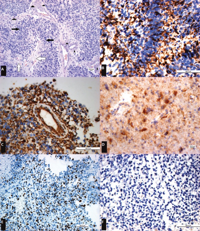
Photomicrographs of the lesion. A: [original magnification, ×200; hematoxylin-eosin stain] demonstrate regions of necrosis (white arrows) surrounded by rings of pseudopalisading tumor cells (black arrows), which are in turn surrounded by abundant microvascular hyperplasia (arrowheads). B: [original magnification, ×400; glial fibrillary acidic protein (GFAP) stain] demonstrate diffuse positivity. C: [original magnification, ×400; Vimentin stain] demonstrate strongly positive expression. D: [original magnification, ×400; IDH-1 stain] showed diffuse positive expression. E: [original magnification, ×200; Ki-67 stain] note a high Ki-67 proliferation index with about 30% of the tumor cells showing nuclear staining. F: [original magnification, ×400; neurofilament (NF) stain] showing negative result.