
Fig. 2.
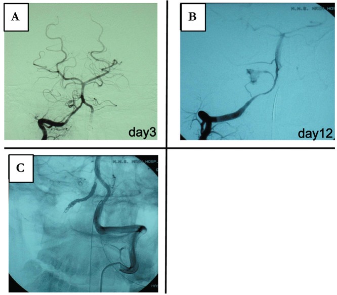
Case 2: Dissection is visible on: A: the right vertebral angiogram of the posterior inferior cerebellar artery (PICA). B: The right vertebral angiogram of the vertebral artery. C: Left vertebral angiogram after embolization of the right vertebral artery.