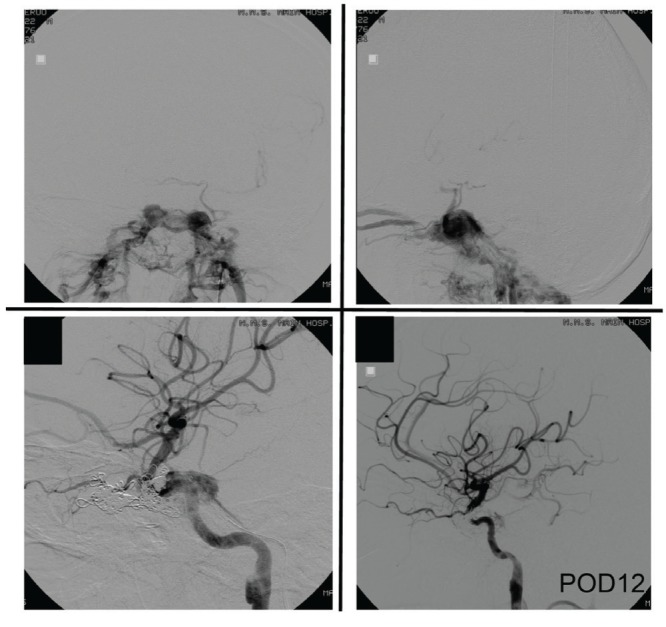
Fig. 3.

Case 3: Left internal carotid angiogram showing traumatic carotid-cavernous fistula with the steal phenomenon (upper panel). Venous reflux is visible in the left and right superior ophthalmic vein and basilar vein. Selective intravenous coil embolization of the fistula was performed. Blood flow in the left internal carotid artery improved after embolization (lower panel).
