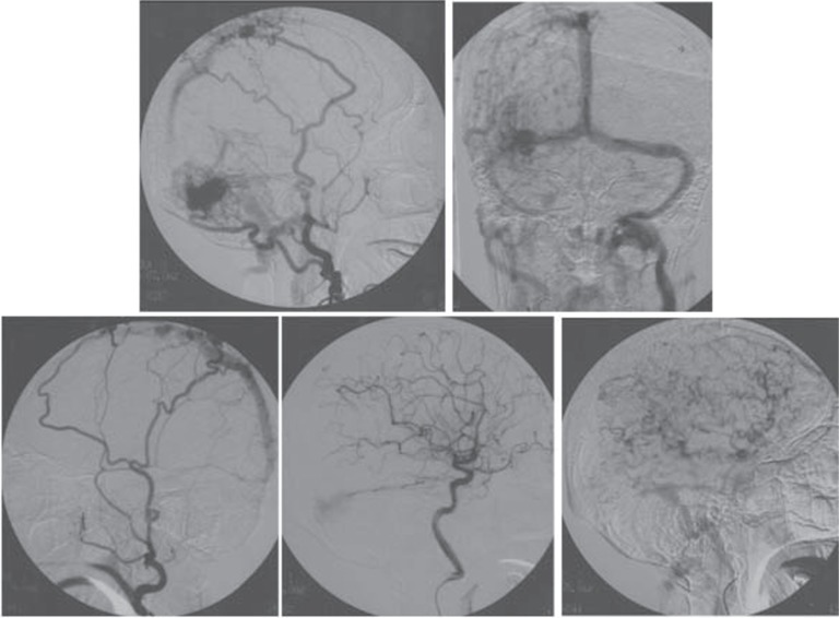
Fig. 2.
Angiograms before surgery. Right external carotid angiograms demonstrating a large dural arteriovenous fistula (AVF) involving the right transverse-sigmoid sinus fed by the right occipital artery, and another dural AVF located in the middle third of the superior sagittal sinus fed by the bilateral middle meningeal and bilateral superficial temporal arteries (upper left, upper right). Left external carotid angiogram demonstrating retrograde venous outflow, draining into the dural AVF of the superior sagittal sinus and cortical veins of the right frontal cortex (lower left). Right internal carotid angiogram demonstrating the dural AVF involving the right transverse-sigmoid sinus fed by the right tentorial artery (lower center). Right internal carotid angiogram (early venous phase) demonstrating markedly severe pseudophlebitic pattern (lower right).

