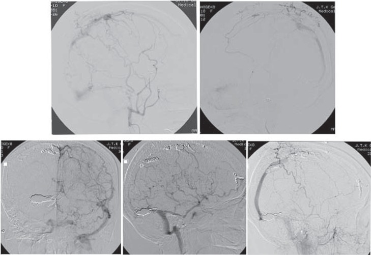
Fig. 4.
Left external carotid angiogram after the second operation demonstrating the right occipital artery occluded by N-butyl-2-cyanoacrylate and Eudragit (upper left). Right external carotid angiogram after the third operation demonstrating that the dural arteriovenous fistula (AVF) of the superior sagittal sinus still remained (upper right). Left internal carotid and right external carotid angiograms after all operations demonstrating that the dural AVF of the transverse-sigmoid sinus, occluded by platinum coils, had completely disappeared, and the dural AVF of the superior sagittal sinus still remained, but the blood flowed anterogradely (lower left, lower right). Right internal carotid angiogram after all operations demonstrating that the pseudophlebitic pattern had completely disappeared (lower center).

