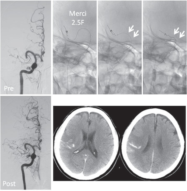
Fig. 2.
Right-sided occlusion in an arch-type horizontal middle cerebral artery (MCA) segment (M1; Case 11). When the Merci retriever is withdrawn, M1 is entirely pulled down and vertical pressure is applied to the microcatheter (white arrows). Subsequently, the retriever loops are stretched. In this case, recanalization was not achieved, and computed tomography performed after the procedures showed the development of subarachnoid hemorrhage.

