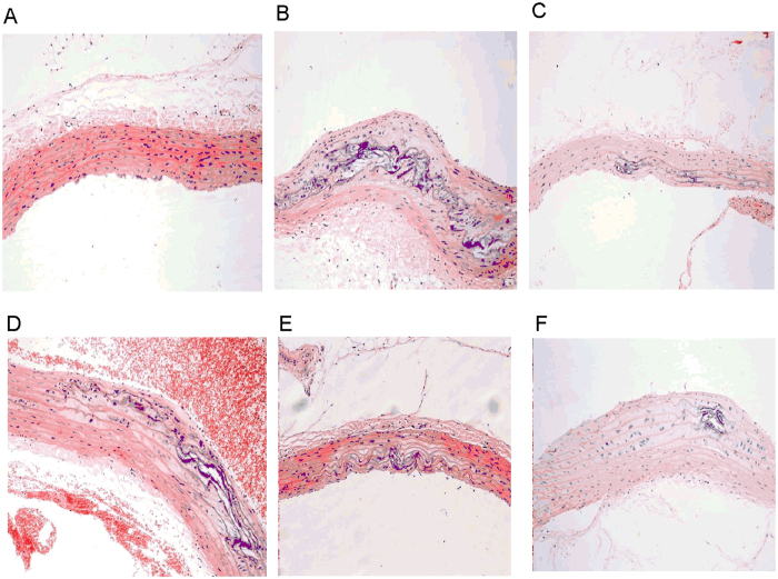
Figure 1. Rat carotid artery sections were subjected to histological examination.
Representative photomicrographs of HE staining are shown. Original magnification: ×200. Normal control (A) showed no impairment of the artery’s integrity and all layers remained intact, whereas atherosclerotic rats exhibited atherosclerotic lesion formation (B). Animals receiving YD (D–F) and atorvastatin (C) intervention showed mild pathological changes compared with normal controls.

