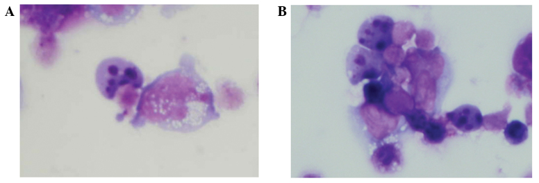
Figure 3.

Detection of apoptosis. (A) Giemsa-stained HBL-2 cells that were incubated with etoposide for 24 h demonstrated an apoptotic morphology (including nuclear fragmentation). (B) Upon neuraminidase pretreatment, the HBL-2 cells also demonstrated an apoptotic morphology.
