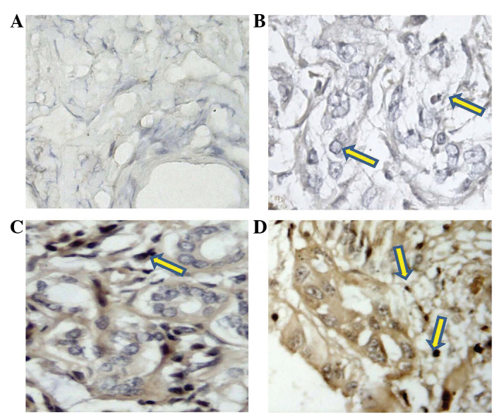
Figure 1.

Immunohistochemical staining for vascular endothelial growth factor C (VEGF-C) in human cholangiocarcinoma. (A) Control specimen; (B) VEGF-C-negative cholangiocarcinoma specimen (arrows indicate VEGF-C was not observed); (C) cholangiocarcinoma specimen with weakly positive VEGF-C expression (arrow); and (D) cholangiocarcinoma specimen with strongly positive VEGF-C expression (arrows).
