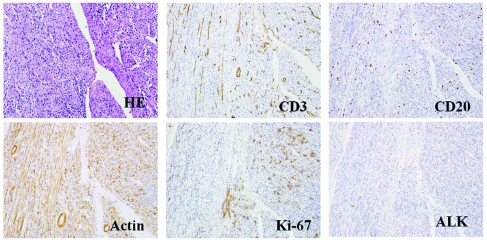
Figure 5.
HE staining and representative histological cross-sections from the recurrent tumor (magnification, x400; microscope, Olympus IX71). Representative histological cross-sections from the recurrent tumors revealed variable myofibroblasts, myxoid stroma and mixed inflammation with lymphocytes, plasma cells and eosinophils. The tumor stained positive for actin, Ki-67, desmin, CD3 and CD20, but negative for ALK and p53. HE, hematoxylin and eosin; ALK, anaplastic lymphoma kinase.

