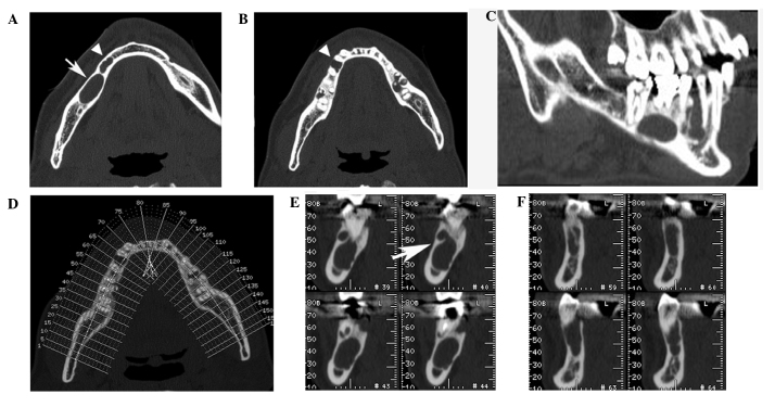
Figure 2.
Computed tomography images of the patient's mandible. (A) An axial view showing the cross-section of the mandible from the patient's foot side. A 15-mm sized, round-style, well-defined cystic lesion was detected on the root apex of lower-right first molar (arrow); in addition, a 12-mm sized, radiolucent lesion was identified in the inter-alveolar septum of the lower-premolars (arrowhead). (B) An axial view demonstrated the root divergency of lower-right premolars (arrowhead). (C) A para-sagittal view revealed the septum inside the ameloblastoma as well as divergency of lower-right premolars. (D) Reference image of the orthogonal reconstruction. Numbers assigned are equivalent to the image number of each orthogonal view. (E) Orthogonal views depicting the keratocystic odontogenic tumor revealed the septum inside the KCOT (arrows). (F) Orthogonal views identifying the ameloblastoma.

