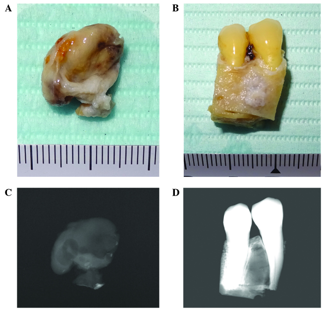
Figure 4.
Soft X-ray images of the excised tumors. Images were captured of (A) the excised KCOT and (B) the ameloblastoma with adjacent teeth and alveolar bone. Soft X-ray images were then captured of (C) the KCOT specimen and (D) the ameloblastoma specimen, which demonstrated details of the internal structures of the lesions. KCOT, keratocystic odontogenic tumor.

