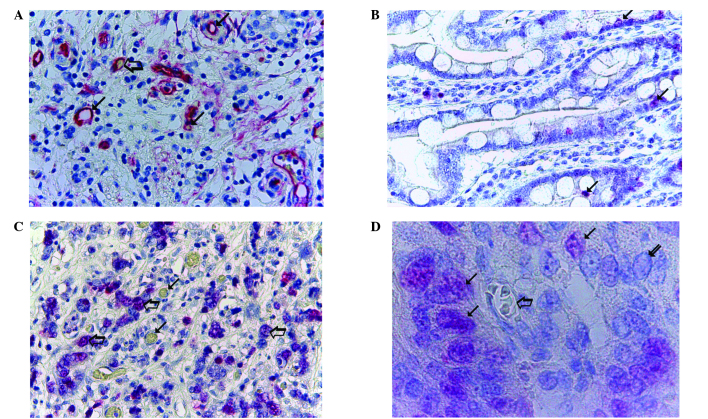
Figure 1.
Morphometrical analysis of MVD and Ki-67 proliferation index in PDAC tissue. (A) Highly vascularised pancreatic ductal adenocarcinoma sample stained with anti-CD-31 antibodies. Numerous red immunostained microvessels are visible: Small arrows, microvessels with a visible lumen; large arrow, microvessel with a red blood cell in its lumen, this acted as an internal positive control (magnification, x400). (B) Well-differentiated and (C) poorly-differentiated PDAC samples stained with anti-Ki-67 antibodies; low and high rates of proliferation are observed, respectively. Arrows, single red-stained proliferating nuclei (magnification, x400). (D) Poorly-differentiated PDAC sample stained with anti-Ki-67 antibodies. Small arrows, single red-stained proliferating nuclei; large arrow, microvessel with several red blood cells in its lumen, this acted as an internal positive control. Red proliferating nuclei are visible near the microvessels (magnification, x1,000, in oil). MVD, microvascular density; PDAC, pancreatic ductal adenocarcinoma.

