
Figure 1.
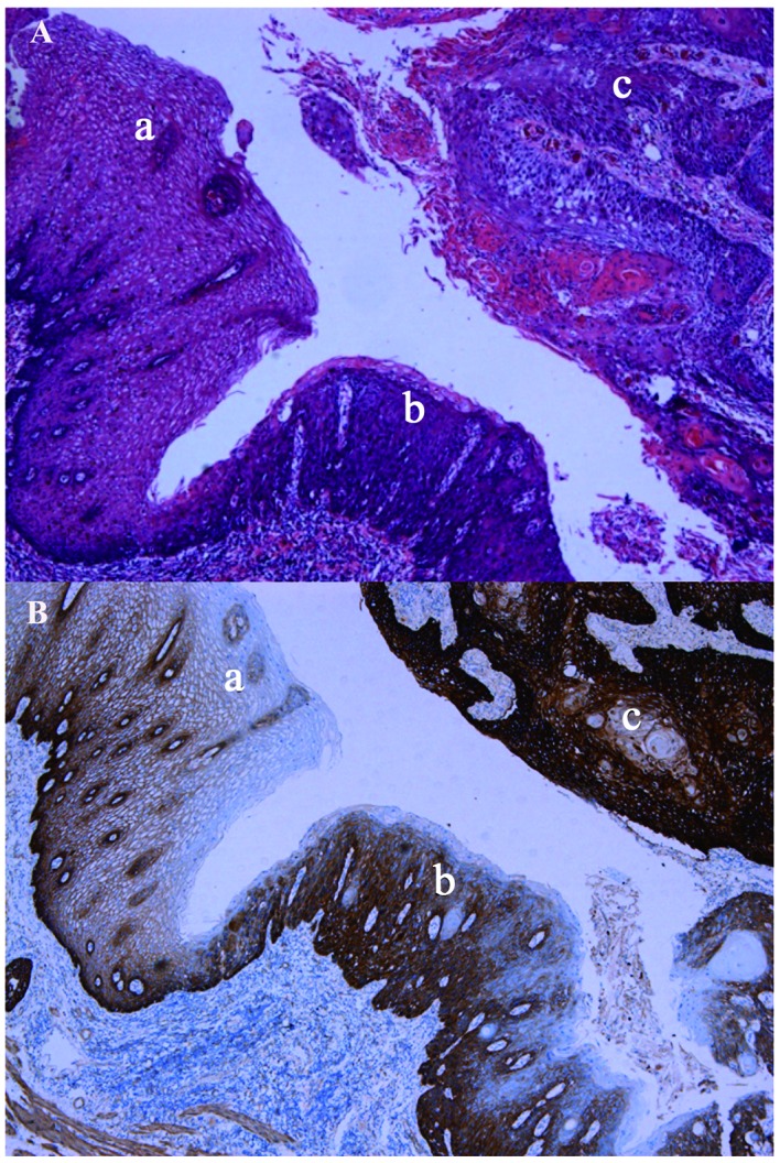
Esophageal squamous cell carcinoma morphology. (A) Hematoxylin and eosin staining and (B) EGFR immunostaining. a, normal esophageal epithelium; b, epithelial atypical hyperplasia; c, tumor tissues. EGFR, epidermal growth factor receptor.
Official websites use .gov
A
.gov website belongs to an official
government organization in the United States.
Secure .gov websites use HTTPS
A lock (
) or https:// means you've safely
connected to the .gov website. Share sensitive
information only on official, secure websites.
Esophageal squamous cell carcinoma morphology. (A) Hematoxylin and eosin staining and (B) EGFR immunostaining. a, normal esophageal epithelium; b, epithelial atypical hyperplasia; c, tumor tissues. EGFR, epidermal growth factor receptor.