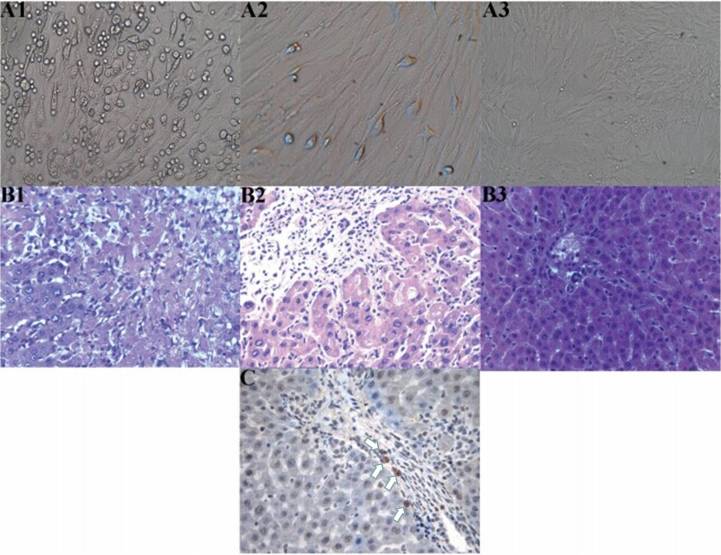
Figure 1.
(A) Cell culture, (B) hepatic histopathological changes and (C) immunohistochemistry showing the location of the mesenchymal stem cells (MSCs). (A) Morphological changes of the MSCs; (A1) Primary culture for three days (magnification, x100) revealed a number of oval-shaped adherent cells and the partial stretching and deformation of cells, presenting as a polygon or clostridium; (A2) First generational passage cell culture (magnification, x200) showed a small number of confluent cells; (A3) Third generational passage cell culture for seven days (magnification, x50) exhibited whirlpool and pectiniform shapes. (B) Hepatic histopathological changes in each group on day 7 after orthotopic liver transplantation (OLT; hematoxylin and eosin stain; magnification, x200); (B1) Group A exhibited evident necrosis of hepatic cells; (B2) Group B exhibited evident inflammatory infiltration, predominantly concentrating in the portal area, without cellular necrosis; (B3) Group C manifested almost no evident inflammatory infiltration. (C) Location of MSCs in the grafts of patients in group C on day 7 following OLT was determined using SRY in situ hybridization (magnification, x400). A number of positive cells (white arrows) can be observed, with the cytoplasm and nucleus stained brown.

