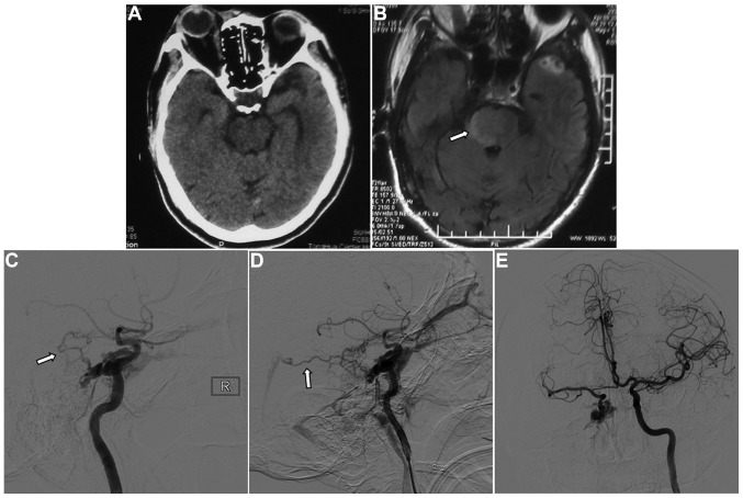
Figure 2.
Images 1 week after the traumatic incident. (A) Computed tomography showed no abnormality. (B) Fluid-attenuated inversion recovery magnetic resonance imaging showed a hyperintense signal in the right side of the pons (arrow). (C and D) Digital subtraction angiography showed that the cavernous sinus drained into the ophthalmic vein, inferior petrosal sinus and clival venous plexus, and the basal vein of Rosenthal potentially enlarging it (arrows). There was no filling of the superior petrosal sinus. (E) Left carotid artery angiography with cross compression of the right carotid artery showed normal collateral circulation in the right carotid artery and no contrast delay in the middle cerebral artery.

