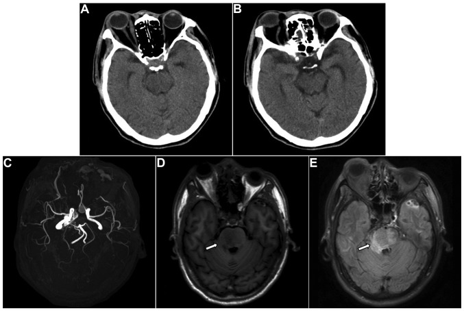
Figure 3.
Radiological images 5 days after diagnosis of carotid-cavernous fistula, during which the patient had left hemiparesis. (A and B) Computed tomography showed no abnormality. (C) Magnetic resonance angiography revealed abnormal enlarged blood vessels in the cavernous sinus. (D) T1W1 and (E) fluid-attenuated inversion recovery magnetic resonance imaging showed aggravation of the brainstem edema (arrows).

