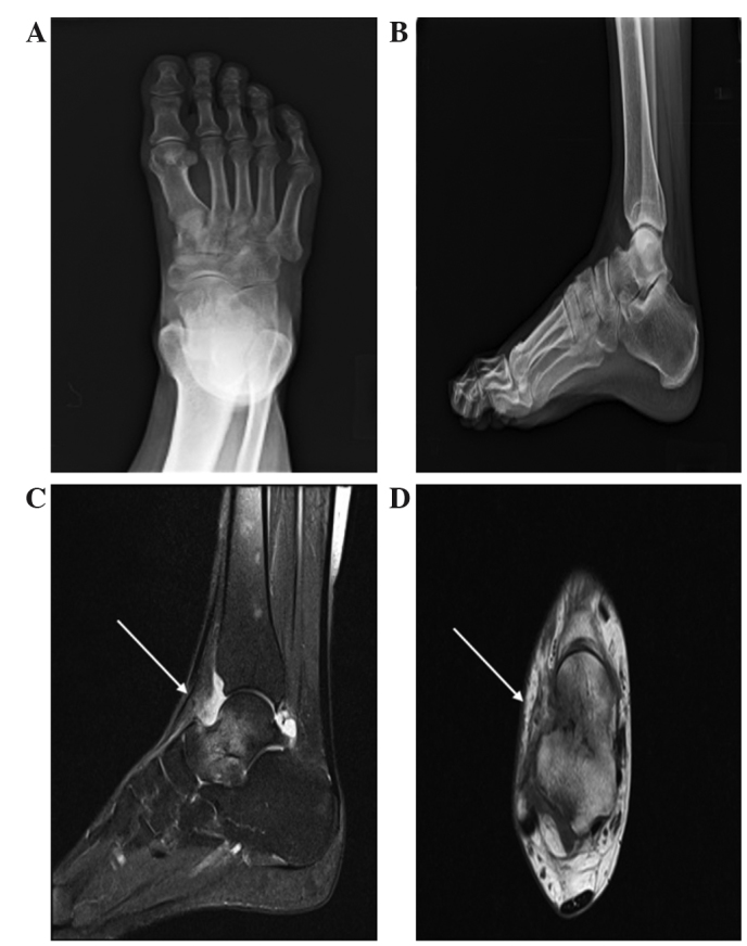
Figure 4.

Plain radiographs and MRI scans in Case II. Plain radiographs of the (A) anterioposterior foot and (B) lateral ankle revealed no specific bony abnormalities. MRI demonstrated inflammatory signal changes on the patients talar neck area with reactive edematous changes (white arrows) on ankle joint of the (C) sagittal and (D) axial scans. MRI, magnetic resonance imaging.
