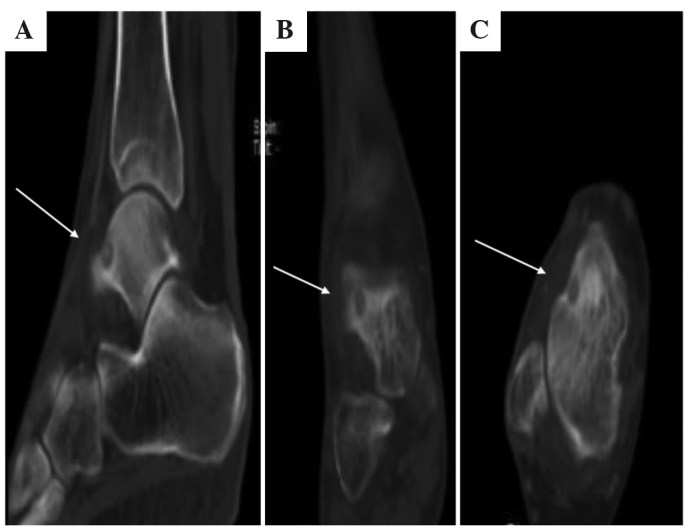
Figure 6.

CT scans in Case II. CT scans of the patients ankle in (A) sagittal, (B) coronal and (C) axial view revealed a small nidus lesion on the talus anteromedial aspect (white arrows), which was diagnosed as osteoid osteoma. CT, computed tomography.
