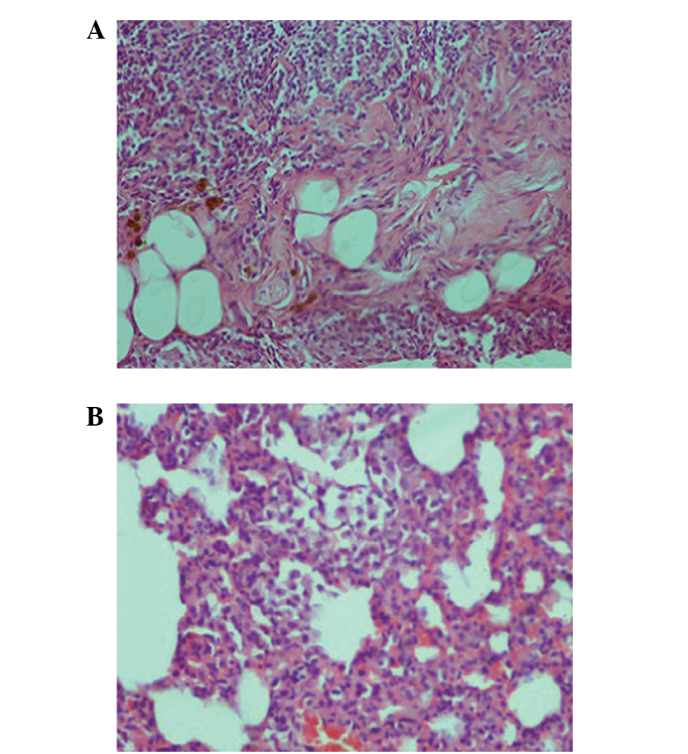
Figure 3.

Histological analysis of the methotrexate group pulmonary tissue. (A) Interstitial lymphocytic inflammation and interstitial fibrosis were observed in the pulmonary tissue (hematoxylin and eosin; magnification, x100). (B) Type 2 pneumocyte hyperplasia was also observed in the tissue (magnification, x200).
