
Figure1.
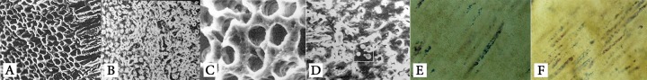
SEM images showing removal (A) and presence (B) of smear layer, bacterial growth in specimens without (C) and with (D) smear layer, microbial growth controlled by Gram staining in specimen without (E) and with (F) smear layer (400× magnification