
Figure 6.
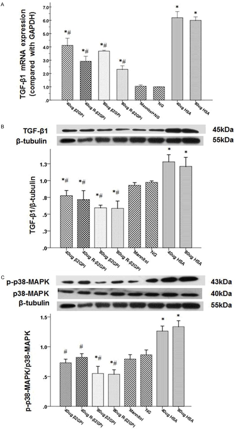
Treatment with β2GPI and reduced β2GPI inhibited the activation of TGF-β1 and p38 MAPK in high glucose-iduced RMs. A and B. Quantification of TGF-β1 expression in high glucose-induced RMCs was performed using quantitative real-time RT-PCR and western blot. The mRNA and protein expression of TGF-β1 were significantly increased after high glucose stimulation and were significantly decreased with a high-dose of β2GPI and reduced β2GPI compared witha ow-dose. C. Quantification of p38MAPK and phospho-p38 MAPK expression in high glucose-induced RMCs was performed using western blot. The phosphorylation level of p38 MAPK significantly increased after high glucose stimulation, whereas β2GPI and reduced β2GPI treatment inhibited p38 MAPK phosphorylation. Data are expressed as the mean ± S.E.M. (n=8 for each group). *P<0.05 v. N group, #P<0.05 vs. DM group. N: normal control; DM: diabetic model control; β2GPI: 20 μg β2GPI; reduced β2GPI: 20 μg reduced β2GPI.