
Figure 6.
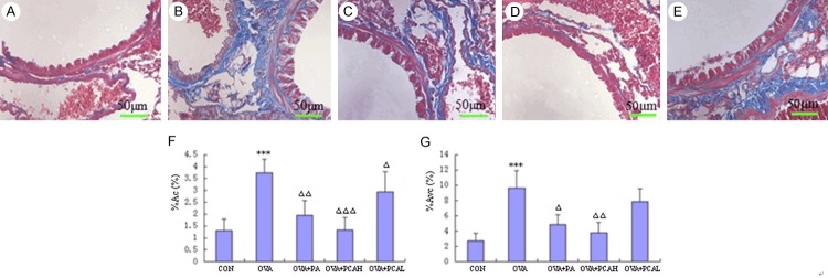
Manson staining (400×) and morphometric measurements of target airway in BABL/c mice. (A) Control group: less airway and perivascular collagen deposition, no or rare collagen deposition in the basement under the plexiform layer. (B) OVA group: a lot of collagen deposition in the airway and vascular smooth muscle surroundings, a lot of collagen deposition in the reticular layer under the airway basal membrane and interstitial lung, the entire field of vision was blue and purple. (C) OVA + PA group: airway and perivascular collagen deposition was significantly reduced compared with OVA group, and only a small amount of collagen fibers in the reticular layer under basement membrane and interstitial lung. (D) OVA + PCAH group: only a few collagen deposition in the entire field of vision, and no difference with the control group. (E) OVA + PCAL group: more airways and perivascular collagen deposition, more obvious interstitial lung collagen deposition. Quantitative and positioning study of morphometry of lung in Manson staining is shown in (F) (the collagen coagulation around bronchial, %ACO) and (G) (the collagen coagulation around vascular, % Avc). N = 10, ***P < 0.001 compared to COM groups. ΔP < 0.05, ΔΔP < 0.01, ΔΔΔP < 0.001 compared to OVA groups.