
Figure 2.
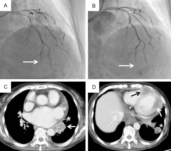
(A, B) Coronary angiography (CAG) and (C, D) contrast-enhanced chest computed tomography (CT) findings. (A) CAG on admission showed occlusion of the distal left anterior descending coronary artery (white arrow). (B) CAG after plain old balloon angioplasty showed revascularization with persistent residual stenosis (white arrow). (C) Contrast-enhanced CT on day 7 showed an irregularly shaped mass in the left lower lobe of the lung adjacent to the left atrium (white arrow). Part of the pericardium was thickened (asterisk). (D) Myocardial masses were present in the apex and intraventricular septum (black arrow) and in the left lateral wall (white arrow). An intrapulmonary metastasis is also visible (asterisk).