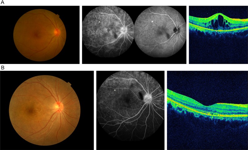
Figure 1.

A. Disc hyperfluorescence, retinal staining, macular edema, and “leopard spots” (arrowhead) were present simultaneously on fundus fluorescein angiography (FFA) of patient 10. “Persistent dark dots” (asterisk) were present on indocyanine green angiography (ICGA). The inner segment/outer segment junction (IS/OS) loss (arrow) and cystoids macular edema (CME) were seen on spectral domain optical coherence tomography (SD-OCT). B. Six months after being treated with a neurosyphilis regimen, the FFA had improved significantly, and the “leopard spots” had disappeared (arrowhead). The IS/OS junction was mostly restored on SD-OCT (arrow), except for a slight disruption, and the CME was eliminated completely.
