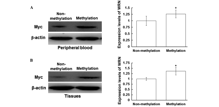
Figure 3.
Expression levels of Myc in the peripheral blood and tissues. Total proteins from the peripheral blood and tissue samples were harvested, separated by 12% SDS-PAGE and analyzed with western blot analysis, where β-actin was used as a loading control. Protein expression levels of Myc in the (A) peripheral blood and (B) tissues were detected using a rabbit anti-human Myc polyclonal antibody (1:500). Left panel, representative western blot; right panel, quantitative assessment of the western blot analysis results. *P<0.05, vs. non-methylation group. WRN, Werner syndrome protein.

