Fig. 3.
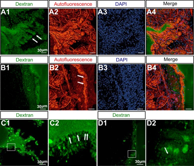
Fine structure of E. virescens electrocytes. A: 3D reconstruction of membrane papillae within a single invagination from serial confocal scans through a fixed 100-μm-thick section of EO tissue. A1 shows the intracellular structure identified by injection of Alexa Fluor 594 dextran (10,000 MW). Arrows point to the spines projecting from membrane papillae. In A2, tissue autofluorescence reveals that the spines shown in A1 are wrapped by capillaries. DAPI stains the multiple nuclei in A3. Channel images A1–A3 are opaquely superimposed in A4. B: 3D reconstruction of the junction between adjacent electrocytes from serial confocal scans through a 100-μm-thick section of fixed EO tissue. B1 shows the anterior face of the electrocyte injected with Alexa Fluor 594 dextran. Blood vessels occupy the space between adjacent electrocytes in B2, and arrows indicate the capillaries penetrating the anterior face. DAPI labels nuclei in B3. Channel images B1–B3 are merged in B4. C: the posterior-face membrane is mostly occupied by vesicles. C1 is an optical section from the serial confocal scans of the posterior face shown in A1. The region outlined in white in C1 is enlarged in C2, where arrows indicate vesicles. D: few vesicles are found on the surface of the anterior face. D1 is an optical section from the serial confocal scans of the anterior face shown in B1. The region outlined in white in D1 is enlarged in D2. Green circular structures in C and D are nuclei. Arrows indicate vesicles.

