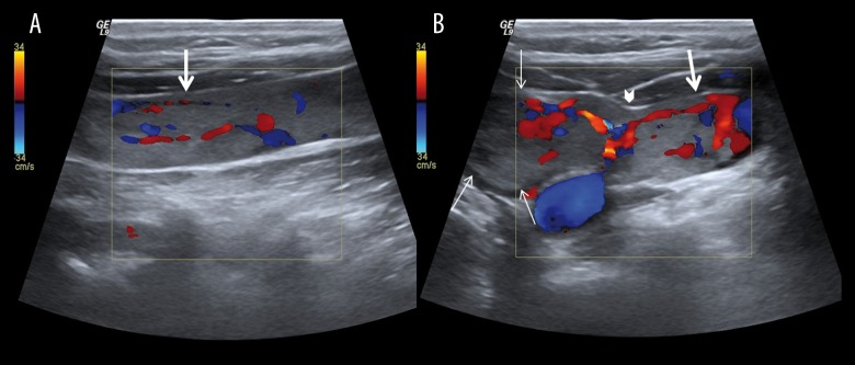
Figure 2.
Color Doppler ultrasound of neck; (A) the longitudinal image of the left internal jugular vein (IJV) shows the hyperechoic filling defect and vascularization of tumour thrombus (thick arrow). (B) the transvers image of the vascularization of thyroid mass (thin arrow) and the invasion region (arrow head).

