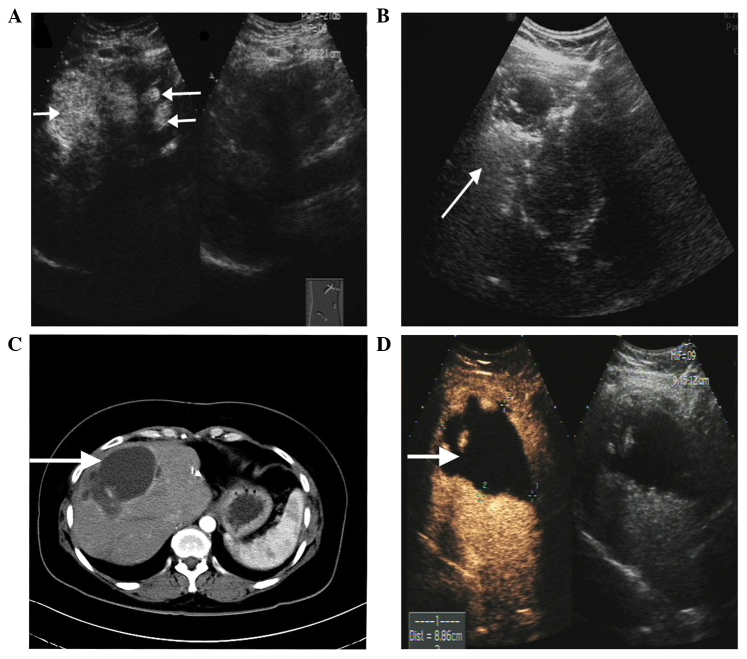
Figure 4.
(A) Preoperative contrast-enhanced ultrasonography revealed multiple hyperechoic tumors in the right liver (largest, 78×53 mm). (B) Ultrasonography immediately following radiofrequency ablation showed the tumor as a hyperechoic region. (C) Abdominal enhanced computed tomography revealed that the initial liver tumors had been replaced by non-enhanced regions (i.e. ablation zones). (D) Contrast-enhanced ultrasonography revealed multiple contrast agent filling defects in the right liver. No liver metastases were observed.

