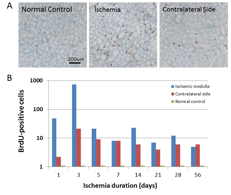
Figure 2 :

BrdU-positive cells were significantly increased in the adult kidney after renal ischemia-reperfusion injury. A. BrdU-positive cells were barely detectable in the sham-operated rats (left panel: lower-magnification view). However, BrdU-positive cells (brown color) in the renal tubules were dramatically increased after transient renal ischemia (middle panel: low-magnification view). BrdU-positive cells in the contralateral side were also increased significantly to compare to that in the normal control, though it is lower than that in the transient renal ischemia. Representative microphotographs of a kidney section were obtained from 3 days after ischemia. Hematoxylin was used for nuclear staining. B. Quantitative analysis of BrdU-positive cells in the renal medulla with normal control, transient renal ischemia and contralateral side. Data shown are mean ± SD. n = 6–8 per group.
