Fig. 3.

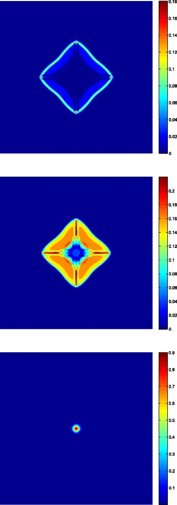
Three layers fully developed distribution of the tumor at time t=270[12 h]. Spatial distribution of proliferative cells (up), quiescent cells (middle) and necrotic cells (down)

Three layers fully developed distribution of the tumor at time t=270[12 h]. Spatial distribution of proliferative cells (up), quiescent cells (middle) and necrotic cells (down)