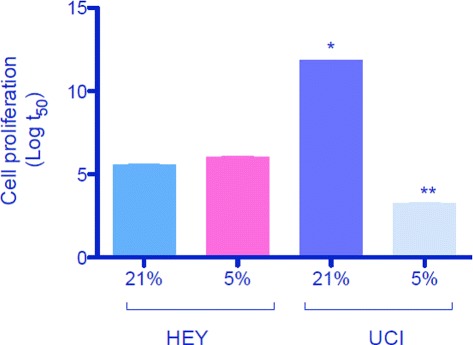
Fig. 4.

Different cell proliferation in ovarian cancer cells. Two cell lines derived from human ovarian cancers were used (HEY and UCI). Cells were cultured under 21 % and 5 % oxygen during 0, 3, 6, 12 and 24 hours. Cell proliferation was analyzed by bromouridine incorporation as previously reported [58]. Data is presented as the logarithm of t 50 (replication time) ± SEM. N=4 per group and analyzed time. * p<0.05 vs HEY at 21 % oxygen. ** p<0.05 vs UCI at 21 % oxygen
