
Fig. 2.
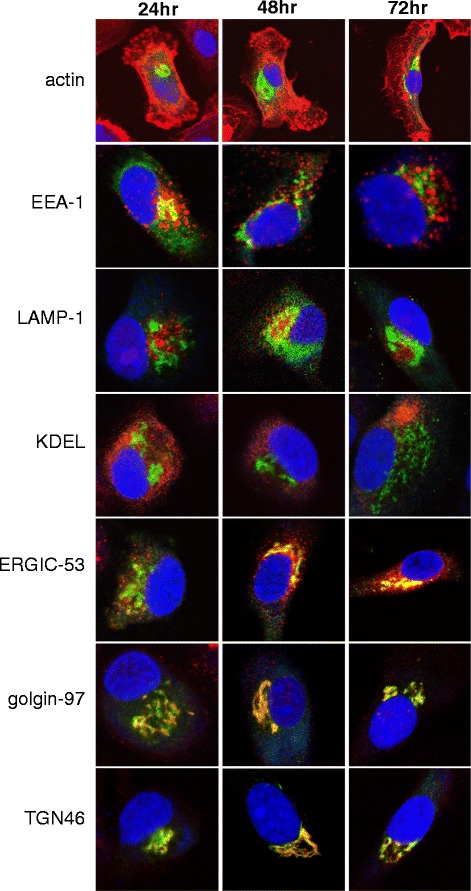
Intracellular trafficking of adenovirally-expressed AFP. DC were transduced with AdVhAFP at MOI 2000 for 3 hr, and then cultured in DC media for 24, 48, or 72 hr. Cells were then fixed and stained for AFP (green), actin, EEA-1 (early endosomes), LAMP-1 (late endosomes/lysosomes), KDEL (endoplasmic reticulum/ER), ERGIC-53 (ER-Golgi intermediate complex), golgin-97 (Golgi), and TGN46 (trans-Golgi network) (all in red), as described in Materials and Methods. All images are representative of three independent experiments performed and were taken using a 63x objective