
Fig. 5.
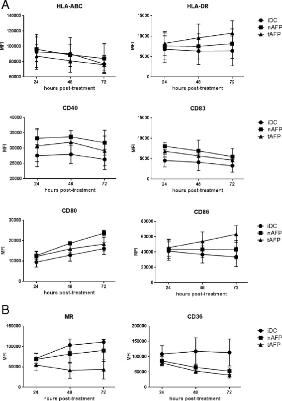
Phenotype of AFP-loaded DC. a and (b) Immature DC (iDC) from healthy donors (n = 3) were left untreated or cultured with nAFP and tAFP (10 μg/ml) in DC media for 24, 48, or 72 hr. Cells were stained for (a) antigen presentation and costimulatory markers and (b) endocytic receptors, and analyzed by flow cytometry. Mean fluorescence intensity (MFI) is reported as the mean ± SD