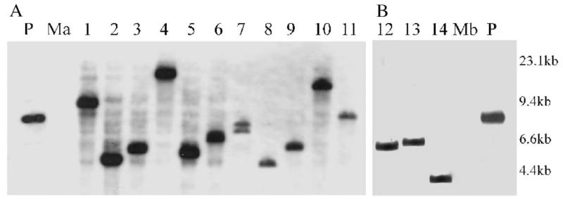
Fig. 2.
Southern hybridization analysis of transformant DNA of M. agalactiae (A, lanes 1–11) and M. bovis (B, lanes 12–14) double-digested with BamHI/EcoRI and probed with a DIG-labeled fragment corresponding to the 400-bp region within the HindIII fragment of Tn4001. Digested DNA corresponding to non-transformed M. agalactiae (Ma) and non-transformed M. bovis (Mb) served as negative controls and pISM2062 (P) as positive control. DNA size standards are indicated in the right margin.

