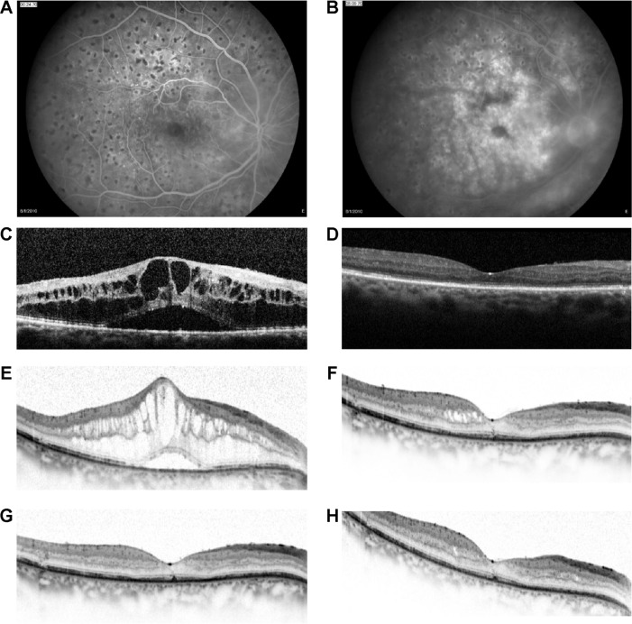
Figure 4.
A 65-year-old female with regressed proliferative diabetic retinopathy in the right eye previously treated with three monthly injections of intravitreal bevacizumab.
Notes: Early- and late-phase fluorescein angiograms reveal significant leakage, with accumulation in a cystoid pattern, and multiple-scatter scars (A and B). Optical coherence tomography images indicate a decrease in central macular thickness from a baseline (post-bevacizumab) level of 846 µm (BCVA 6/30) (C) to a trough of 209 µm (BCVA 6/15) at 11 weeks after the first dexamethasone implant injection (D), with reversal of effect occurring by week 20 (CMT 752 µm; BCVA 6/15) (E). Consistent, marked reductions in central macular thickness to trough levels of 222 µm (BCVA 6/12), 209 µm (BCVA 6/30), and 235 µm (BCVA 6/15) were recorded 9–12 weeks after the second, third, and fourth dexamethasone implant injections, respectively (F–H). Images courtesy of Dr A Loewenstein.
Abbreviation: BCVA, best-corrected visual acuity.

