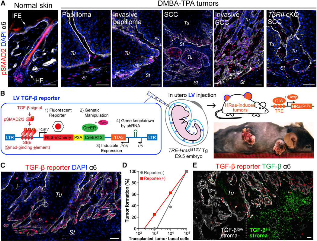
Figure 1. Lentiviral TGF-β Reporter System for Probing Malignant Transformation In Vivo.
(A) pSMAD2 immunolocalization in normal mouse skin and at different stages of DMBA-TPA-induced malignant progression to SCC. Integrin α6 denotes the boundary of tumor epithelia (Tu) and stroma (St). IFE: interfollicular epidermis, HF: hair follicle.
(B) Schematic of LV-mediated in vivo TGF-β reporter and KO/KD system. NLS-mCherry and CreER are under the control of TGF-β signaling. shRNA and rtTA3 transcription factor are under constitutive promoter regulation. LV transduction of surface epithelium of live E9.5 TetO-HrasG12V X Rosa-YFP embryos was achieved by in utero ultrasound-guided microinjection into the amniotic sac. Doxy-induction of HRasG12V initiates tumorigenesis. When desired, CreER is activated by Tam to induce recombination-dependent Rosa-YFP.
(C) Epifluorescence detection of TGF-β–pSMAD2 signaling in HRasG12V SCC.
(D) Limit-dilution orthotopic transplantation of primary tumor basal cells ± TGF-β reporter activity (103 and 104 cells; n=8, 105 cells; n=3).
(E) Epifluorescent TGF-β reporter activity with pan-anti-TGF-β and anti-α6 immunofluorescence shows that basal tumor cells with high TGF-β reporter activity are juxtaposed to stroma with high TGF-β (right). Note heterogeneity demarcated by vertical dotted line.
Scale bars, 50 µm. See also Figure S1.

