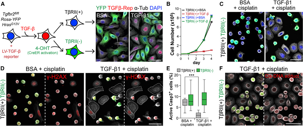
Figure 3. Active TGF-β Signaling in Basal Tumor Cells Increases Their Protection against Cisplatin-Induced Apoptosis.
(A) Experimental scheme to derive isogenic HRasG12V-expressing YFPnegTβRII+ and YFP+TβRIIneg MKs from 10 cultures. (Right) Epifluorescence of cultures ± TGF-β1. Note that only YFPnegTβRII+ cells are TGF-β reporter+ (mCherry+).
(B) Growth curves of TβRII+ and TβRIIneg cells ± 100pM TGF-β1. Data are mean ± SEM.
(C) Immunofluorescence of BSA- or TGF-β1-pretreated (36 hr) YFPnegTβRII+ and YFP+TβRIIneg MKs treated 10 hr with cisplatin. Note that YFP+TβRIIneg MKs frequently displayed signs of apoptotic rounding.
(D) γ-H2AX detection of DNA double-strand breaks induced by cisplatin treatment. Note that both YFPnegTβRII+ and YFP+TβRIIneg MKs show γ-H2AX signal in control, but TGF-β1 selectively spares TβRII+ cells, which remain spread.
(E) Quantifications of AcCasp3+ cells in the same experiment as in (C) and (D) (n=15 microscopic image fields). Data are box-and-whisker plots.
(F) Immunodetection of adduct formed between cisplatin and DNA. Note that YFP+TβRIIneg cells have more cisplatin-modified DNA (red). Anti-tubulin (white). Scale bars, 50 µm.

