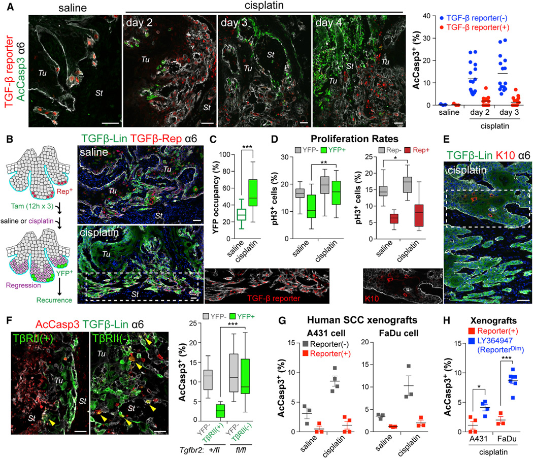
Figure 4. TGF-β-Responding SCC-SCs Show Enhanced Drug Resistance In Vivo.
(A) Immunodetection of AcCasp3 (green) and TGF-β reporter (red) in tumors from mice administrated with saline or cisplatin. (Right) Quantifications revealing that TGF-β signaling protected basal tumor cells from cisplatin-induced apoptosis. (3 tumors analyzed; >15 microscopic image fields per tumor).
(B) Experimental scheme and representative examples of lineage tracing to monitor the fate of the TGF-β reporter+ subset of basal tumor cells after cisplatin treatment. (Right) Note that resistant SCCs are largely contributed by TGF-β-responding cells that survived cisplatin.
(C) Quantifications show that the % of YFP+ basal tumor cells increases after cisplatin, suggestive of their preferential survival.
(D) Quantifications of basal tumor cells in G2/M phase (phospho-H3+) that are YFPneg vs YFP+ (left) and reporterneg vs reporter+ (right). Note: although proliferation of basal tumor cells that resist cisplatin is generally elevated, TGF-β-responding SCC-SCs are still slower-cycling.
(E) Recurring tumor clones that resist cisplatin are largely YFP+ and express little K10, consistent with their derivation from TGF-β reporter+ basal cells.
(F) Quantification of apoptotic cells in cisplatin-treated tumors from LV-transduced Tgfbr2+/fl or Tgfbr2fl/fl mice treated as in (B). Note enhanced survival of TβRII+ progenies.
(G and H) Quantification of AcCasp3+ cells after cisplatin treatment of xenografts of (G) TGF-β reporter transduced human SCC cells (reporter+ vs reporterneg), and (H) xenografts pre-treated ± LY364947 (reporter+ vs reporterdim) (n>3).
Data are box-and-whisker plots (C–F) and mean ± SEM (G and H). Scale bars, 50 µm. See also Figure S3.

