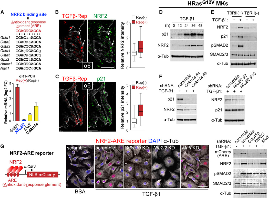
Figure 6. TGF-β Target p21 Is Required for NRF2-Dependent Activation of Antioxidant Genes.
(A) Nucleotide sequence of NRF2 binding motifs within the 5′-upstream region of Gst and other NRF2 target genes. Nucleotides in capital letters are those shared by the antioxidant response element (ARE) consensus sequence. (bottom) qRT-PCR analysis of in vivo tumor basal cell RNA samples. Data are mean ± SEM.
(B and C) Co-expression of NRF2 or p21 (green) and TGF-β reporter (NLS-mCherry) at tumor-stroma interface of TβRII+ tumor sections. Fluorescent intensities of NRF2 and p21 staining in TGF-β reporter+ and reporterneg cells were quantified (NRF2: n=78 and 57 cells, p21: n=101 and 71 cells). Data are box-and-whisker plots.
(D) Immunoblotting of lysates prepared from HRasG12V-overexpressing TβRII+ 10MKs stimulated with TGF-β1 for indicated times.
(E) Immunoblotting of lysates prepared from HRasG12V-overexpressing TβRII+ and TβRIIneg 10MKs stimulated with TGF-β1 for 36 hr.
(F) Immunoblotting of lysates prepared from 36 hr TGF-β1-treated HRasG12V-overexpressing 10MKs transduced with scramble, Cdkn1a or Nfe2l2 shRNAs.
(G) LV NRF2-ARE reporter. (Right) Immunofluorescence and immunoblots of NRF2-reporter transduced HRasG12V-induced MKs expressing scramble (control), Cdkn1a, Nfe2l2, or Maff (control) shRNAs ± TGF-β1 stimulation (36 hr). Note that ARE-reporter activity is abolished upon Cdkn1a or Nfe2l2 but not control KD. Scale bars, 50 µm. See also Figure S5.

